erythrocyte rosette test
In experiments with simulated fetomaternal hemorrhage all 79 samples containing from 25 to 70 ml of fetal whole blood were positive according to the erythrocyte rosette test. It is Erythrocyte-Rosette Formation test.

Neutrophil Erythrocyte Rosettes In Autoimmune Hemolytic Anemia Schulman 2013 American Journal Of Hematology Wiley Online Library
Presence of erythrocyte rosetting mostly by monocytes and massive erythrophagocytosis in peripheral smear has been reported very.

. Fetuin glycopeptide blocks rosette formation when added to a suspension of human lymphocytes and sheep erythrocytes or when preincubated with human lymphocytes but not when preincubated with sheep erythrocytes. A new test termed the human allogeneic rosette test HART is reported for the detection of small amounts of erythrocyte autoantibodies. The Rosette test is performed on postpartum maternal blood to estimate the volume of fetal-maternal hemorrhage in case of an Rh negative mother and an Rh positive child.
A new test termed the human allogeneic rosette test HART is reported for the detection of small amounts of erythrocyte autoantibodies. If the rosette test is positive follow-up testing is done to quantitate the FMH eg a Kleihauer-Betke acid elution test or flow cytometry. The red cell eluate was further tested with the euroimmune anti-sars-cov-2 spike s1 elisa and was negative suggesting that the neutrophil-erythrocyte rosettes seen were unlikely due to anti s1 igg immunoglobulins.
This estimate in turn also estimates the required. Donath Landsteiner test DL is the gold standard diagnostic test. Immunoglobulin levels were found to be largely normal although several patients were clearly hypogammaglobulinemic.
The red blood cells surrounding the cell form the petal while the central cell forms the stigma of the flower shape. Applying the Du test to the same samples resulted in a 30 false negative rate at the level of a. In experiments with simulated fetomaternal hemorrhage all 79 samples containing from 25 to 70 ml of fetal whole blood were positive according to the erythrocyte rosette test.
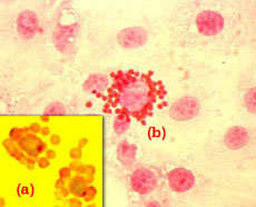
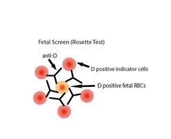
Erythrocyte rosetting or E-rosetting is a phenomenon seen through a microscope where red blood cells erythrocytes are arranged around a central cell to form a cluster that looks like a flower. The anti-D binds to D-positive fetal RBCs and when indicator D-positive RBCs are added rosettes are formed. Three types of rosette techniques have been developed and used experimentally.
Enzyme treated indicator cells are added only binding to the fetal cells that were present and sensitized resulting in a process called erythrocyte rosetting or E-rosetting. Erythrocyte-Rosette Formation test - How is Erythrocyte-Rosette Formation test abbreviated. Rosette of sheep red blood cells around the mud catfish lymphocytes were observed in all experimental groups.
It compares the percentage of rosettes formed around lymphocytes from normal subjects when either red cells from patients are added or autologous or allogeneic normal red cells. ERFT - Erythrocyte-Rosette Formation test. Erythrocyte Rosette Test T-like lymphocytes Mud catfish.
The cell mixtures were centrifuged 1000 rpm 5 minutes and pellets were resuspended in MEM. This method has an FMH detection limit of about 10 mL. There existed a laboratory test for rosette formation between patient erythrocytes and sheep monocytes the monocyte monolayer assay that had many limitations but greater predictive value for clinically significant haemolysis than the standard Coombs test.
Rosette Test Fetal RBC Screen Qualitative The rosette test demonstrates the number of D-positive cells in a D-negative suspension using an anti-D reagent. Erythrocyte-Rosette Formation test listed as ERFT. Applying the Du test to the same samples resulted in a 30 false negative rate at the level of a.
Citation neededRosette test for Rh factor. The rosette test is a screening test for FMH that detects fetal D red cells in maternal Rh negative blood. Looking for abbreviations of ERFT.
A 02 ml aliquot of cell suspension 110 6 lymphocytes in MEM of unvaccinated animals was mixed with 02 mL of erythrocytes 1210 6 cells and incubated at room temperature 30C for 30 minutes. At that point T-cells were identified by sheep erythrocyte rosetting and the patients were found to have a broad range of T-cell counts in the peripheral blood. This estimate in turn also estimates the required amount of RhoD immune globulin to administer.
The results suggest that the mouse erythrocyte receptor is an early marker in B cell differentiation and that receptor expression by individual cells may depend on the. The numbers of rosette formed were significantly higher p. Jefferis Use of antibody-coated red cells for the sensitive detection of antigen and in rosette tests for cells bearing surface immunoglobulins.
In this test a sample of maternal blood is incubated with RhoD immune globulin which will bind to any fetal. Erythrocyte rosette formation assay. Three types of rosette techniques have been developed and used experimentally.
Indeed a defunct laboratory test for rosette formation between patient erythrocytes and sheep monocytes the monocyte monolayer assay had many limitations but greater predictive value for clinically significant hemolysis than the standard Coombs test 1 5. The indicator cells will be at the center of the rosette while the fetal RBCs will be clustered around the edges like petals on a flower. Studies of the binding of 3H fetuin glycopeptide to normal lymphocytes demonstrate 75 x 106 saturable binding sites per cell.
Use of the erythrocyte rosette test to screen for excessive fetomaternal hemorrhage in Rh-negative women Abstract The possibility of Rh immune globulin failure exists when a fetomaternal hemorrhage exceeds 25 to 30 ml of whole blood and only one 300 micrograms vial of Rh immune globulin is administered. It compares the percentage of rosettes formed around lymphocytes from normal subjects when either red cells from patients are added or autologous or allogeneic normal red cells. The Rosette test is performed on postpartum maternal blood to estimate the volume of fetal-maternal hemorrhage in case of an Rh negative mother and an Rh positive child.
Request PDF The Application of Erythrocyte Rosette Test to Characterize T-Like Lymphocytes in the Mud Catfish Clarias gariepinus The Erythrocyte rosette test a technique used to. 2 months after recovery from covid-19 the repeat pbf showed resolution of neutrophil-erythrocyte rosettes with a negative direct. This formation occurs due to an immunological reaction between an epitope.
How Are The Cells Studied

The Rosette Screen One Drop Of The Manufacturer S Reagentreduced Download Scientific Diagram
E Rosette Test Liberal Dictionary
2

The Rosette Screen One Drop Of The Manufacturer S Reagentreduced Download Scientific Diagram

Orange Liquid Isolation Of Rosette Cells Teaching Kit For Laboratory Id 9559666633
2

Test 6 Specific Cellular Immune Function Assay Separation Of Mononuclear Cells From Human Peripheral Bloodseparation Of Mononuclear Cells From Human Ppt Download

A Normal Viable Lymphocytes Which Have Formed Rosettes With Download Scientific Diagram

Lecture Notes In Medical Technology Lecture 9 Hemolytic Disease Of The Newborn
Nanomalaria Institute For Bioengineering Of Catalonia
Frontiers Naturally Acquired Humoral Immunity Against Plasmodium Falciparum Malaria

Lymphocyte From Pbl Showing Rosette Formation With Neuraminidase Download Scientific Diagram
Spontaneous Sheep Erythrocyte Rosettes Are Positive In This Download Scientific Diagram

Rosette Kleihauer Betke Tests Overview Procedure What Is A Rosette Study Com
Leukocyte Erythrocyte Rosettes Blood Academy

Plasmodium Falciparum Rosetting Protects Schizonts Against Artemisinin Ebiomedicine

Leukocyte Erythrocyte Rosettes Blood Academy

Stevor Is A Plasmodium Falciparum Erythrocyte Binding Protein That Mediates Merozoite Invasion And Rosetting Cell Host Microbe


Comments
Post a Comment